Computed Tomography - CT/CAT Scan
Exam Explanation
 CT uses special x-ray equipment to obtain image data from different angles around the body. The computer takes the data and creates a visual image of each slice of information. The Radiologist is able to review the slices of information in sequence, which creates a two or three dimensional image of the inside of your body. CT imaging is particularly useful because it can show several types of tissue: lung, bone, soft tissue and blood vessels with great clarity. CT helps the Radiologists diagnose problems such as cancers, cardiovascular disease, infectious disease, trauma and musculoskeletal disorders.
Exam Preparation
Tell your physician or the technologist if there is any possibility that you are pregnant or if you have a history of allergies. Prior to the exam a technologist may speak with you on the phone to obtain necessary medical information and discuss instructions to be followed the day of the exam.
During the Exam
After you arrive for your appointment, depending on the area of your body to be scanned, you may need to change into a gown. Some scans may require a contrast agent to help highlight the areas inside your body. This is given in the form of a drink and/or injection. Once you are prepared for your exam, your technologist will help position you on the table of the CT scanner. Usually you will lay face up and the table will move into the large donut shaped scanner. While inside the scanner, you will be able to see your outside surroundings. The technologist will talk with you from the control room where they can see you at all times. It is very important to lay completely still. Periodically, the voice-activated component may speak to you, instructing you to hold your breath for a short period, not swallow, etc. Most CT examinations take about 15-20 minutes.
3D mammogram
 The POWER and PROMISE of 3D MAMMOGRAM is now available at Element Medical Imaging.
Element Medical Imaging is excited to offer 3D mammogram, also referred to as breast tomosynthesis, for breast cancer screening. 3D mammogram produces a three-dimensional view of the breast tissue that helps our Radiologists identify and characterize individual breast structures without the confusion of overlapping tissue.
We believe 3D mammogram will benefit all screening and diagnostic mammogram patients, and is especially valuable for women receiving a baseline screening, those who have dense breast tissue and/or women with a personal history of breast cancer.

3D mammograms can detect 41% more cancers than 2D
Breast cancer screening with 3D is proven to detect an average of 41% more invasive breast cancers compared to 2D alone. The 3D technology gives our Radiologists increased confidence with up to a 40% reduction in recall rates.
The 3D mammogram screening experience is similar to a traditional mammogram. During a 3D exam, multiple, low-dose images of the breast are acquired at different angles. These images are then used to produce a series of one-millimeter thick slices that can be viewed as a 3D reconstruction of the breast.
Early detection means a 98% five-year survival rate
By offering women the latest and more accurate technology in mammography, Element Medical Imaging expects to increase the number of area women who will be routinely screened. Breast cancer is the second leading cause of cancer death among women, exceeded only by lung cancer. Statistics indicate that one in eight women will develop breast cancer sometime in her lifetime. The stage at which breast cancer is detected influences a woman’s chance of survival. If detected early, the five-year survival rate is 98 percent. Almost all insurance plans cover the additional cost for a 3D mammogram.
Services & Procedures
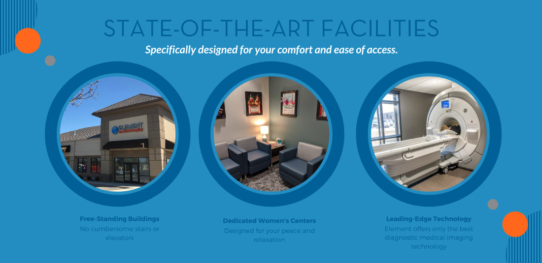
Element Medical Imaging offers a wide array of advanced specialty diagnostic services including MRI, CT, Ultrasound, Mammograms (screening and diagnostic), X-ray, DEXA, flouoroscopy, nuclear medicine studies and more! We offer state-of-the-art equipment in a calm and caring environment. A well-trained, qualified technologist will perform the imaging procedure which is then interpreted by one of our highly-skilled radiologists. Click on an exam listed below to learn more about a specific exam or procedure and any preparation that is needed. Please contact us, if you have any additional questions.
Breast Imaging
- Screening Mammography
- Diagnostic Mammography
- Tomosynthesis (3D Mammography)
- Breast MRI
- Breast Ultrasound
- Needle Biopsy of the Breast
Computed Tomography
Diagnostic X-ray
Fluoroscopy
- General Fluoroscopy Info
- Small Bowel Series
- Upper GI Series
- Hysterosalpingogram
Magnetic Resonance Imaging
Nuclear Medicine
Ultrasound
DEXA (Bone Density)


