


Fertility Ultrasound
Exam Explanation
A fertility ultrasound is a non-invasive imaging modality that uses high-frequency sound waves to create detailed images of the reproductive organs. This exam aids in assessing the health and structure of the uterus, ovaries and fallopian tubes, providing essential information for fertility assessments and treatments.
At Element Medical Imaging, we understand the importance of precise and compassionate care in your fertility journey. Our specialized fertility ultrasound services are designed to provide you and your healthcare provider with detailed insights into your reproductive health.
Common uses of fertility ultrasounds include:
- Follicle Count: Monitoring follicle development and ovulation timing.
- Diagnosing Uterine Abnormalities: Detecting fibroids, polyps or congenital anomalies
- Accessing Ovarian Health: Identifying ovarian cysts and other conditions affecting fertility.
Element Medical Imaging also offers Hysterosalpingogram services.
Exam Preparation
Follicle Count: No preparation required. Bladder must be empty for this exam.
Hysterosonogram: This exam must be performed between days 7-12 of your cycle if you are premenopausal. This exam CANNOT be performed if you are pregnant. It can be performed anytime for post-menopausal patients.
Trans Abdominal (Pelvic): Complete 32oz of water 1 hour prior to your exam time and HOLD. Please do not use the restroom after you start drinking water. Your bladder must be full for this exam.
Trans Abdominal (Pelvic) and Trans Vaginal Ultrasound: Complete 32oz of water 1 hour prior to your exam time and HOLD. Please do not use the restroom after you start drinking water. Your bladder must be full for the Trans Abdominal portion of this exam. You will use the restroom before the Trans Vaginal portion.
During the Exam
Your technologist will discuss your history with you, and you will be positioned face-up on a padded exam table. Depending upon the exam, you may be asked to change into a gown. Your exam will take 30-60 minutes to complete. The radiologist will review your images and send the results to your referring provider.
Breast MRI
What is Breast MRI?
Breast MRI is a sophisticated technology that uses a computer, magnetic field and radio waves instead of x-rays to produce images of the soft tissues in the body. This non-invasive procedure helps Element Medical Imaging’s Radiologists to better evaluate the breast in special circumstances. When used in conjunction with screening and diagnostic mammography, it can provide valuable information for the detection and characterization of breast disease.
Breast MRI has been proven to be more sensitive than any other modality in detecting invasive breast cancer. MRI is superior at demonstrating the size and extent of a breast tumor prior to surgery. In addition, it is beneficial for screening patients at particularly high risk for breast cancer due to genetic predisposition or strong family history, diagnosing breast implant rupture, staging breast cancer and planning treatment. MRI also plays an important role in post-surgical and post-radiation follow-up. In March 2007, the American Cancer Society revised the breast cancer early detection guidelines, recommending annual breast MRI screening for women in the following groups:
- have a known BRCA1 or BRCA2 gene mutation
- have a first-degree relative (mother, father, brother, sister or child) with a BRCA1 or BRCA2 gene mutation, and have not had genetic testing themselves
- have a lifetime risk of breast cancer of about 20% to 25% or greater, according to risk assessment tools that are based mainly on a family history that includes both her mother's and father's side
- had radiation therapy to the chest when they were between the ages of 10 and 30 years
- have a genetic disease such as Li-Fraumeni syndrome, Cowden syndrome, or Bannayan-Riley-Ruvalcaba syndrome, or have one of these syndromes in a first-degree relatives
Women at moderately increased risk (15% to 20% lifetime risk) should talk with their physicians about the benefits and limitations of adding MRI screening to their yearly mammogram. These patient groups include:
- have a lifetime risk of breast cancer of 15% to 20%, according to risk assessment tools that are based mainly on family history (see below)
- have a personal history of breast cancer, ductal carcinoma in situ (DCIS), lobular carcinoma in situ (LCIS), atypical ductal hyperplasia (ADH), or atypical lobular hyperplasia (ALH)
- have extremely dense breasts or unevenly dense breasts when viewed by mammograms
What to expect the day of the exam
The entire appointment will take 45 minutes to an hour. This exam should be scheduled 7-10 days after the start of your menstrual cycle. Correct timing is important to minimize false positive findings that can occur due to hormonal influence on the breast tissue. If you suffer from minor claustrophobia or anxiety, you may want to ask your physician for a mild sedative. Do not take the sedative until you have signed your paperwork. A Breast MRI does not require your breasts to be compressed, so you should not experience discomfort.
When you arrive you will be asked to complete paperwork regarding your history and symptoms. We will escort you into a private dressing room where you can change into a gown and remove all jewelry, since these items contain metal, which disturbs MRI signals. A female technologist will position you for the scan. During the exam, you will lie on your stomach with your arms up over your head and you will enter the machine head first. Avoid eating a large meal prior to the exam. Most patients receive an injection of contrast material called gadolinium during the exam through an intravenous injection. A small intravenous catheter will be placed in your hand or arm. The injection of contrast material is necessary if the MRI is being performed for the diagnosis of breast cancer. It is sometimes not necessary if the sole intent of the study is to evaluate silicone breast implants. Adverse reactions to gadolinium are rare.
You will be asked to lie very still, relax and breathe normally. The MRI scan will only take 30-45 minutes. There are typically no side effects during or after MRI, so you can resume normal activities as soon as your exam is over. It is very important that any prior breast films (mammograms, ultrasound or MRI) be made available to the radiologist for comparison during the interpretation of your MRI scan. If you have had these at a facility other than Element Medical Imaging please bring them with you on the day of your appointment. If you have any of the items listed below, please call 913.469.8998 so we can make arrangements for you before your appointment. Many of these items are contraindications to having an MRI as they are not compatible with the magnetic field present around all MRI machines.
- Cardiac pacemaker
- Artificial heart valve prostheses
- Aneurysm clips
- Eye implants or metal ear implants or any metal implants activated electronically, magnetically or mechanically.
- Copper 7 IUD
- Shrapnel or non-removed bullet
- Pregnancy
- Weight over 350 lbs.
- Claustrophobia
- Any metal puncture(s) or fragment(s) in eye
Nuclear Medicine Scans
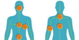
![]() Nuclear Medicine is a subspecialty within the field of radiology. It includes diagnostic imaging studies that demonstrate body anatomy and function. The images are based on the distribution of a radioactive substance given to the patient intravenously. Generally, radiation to the patient is similar to that resulting from standard X-ray examinations. Nuclear medicine images can assist the physician in diagnosing diseases. Tumors, infections and other disorders can be diagnosed by evaluating organ function.
Nuclear Medicine is a subspecialty within the field of radiology. It includes diagnostic imaging studies that demonstrate body anatomy and function. The images are based on the distribution of a radioactive substance given to the patient intravenously. Generally, radiation to the patient is similar to that resulting from standard X-ray examinations. Nuclear medicine images can assist the physician in diagnosing diseases. Tumors, infections and other disorders can be diagnosed by evaluating organ function.
Due to the variation of Nuclear Medicine procedures performed, you will receive exam preparation and instructional information specific to your appointment at the time of scheduling.
Screening and Diagnostic Mammogram
![]() A mammogram is special low-dose x-ray of the breast. At Element Medical Imaging, our Radiologists use the images to detect breast cancer. 3D mammogram has become the gold-standard for breast cancer screening and is one of the most recent advances for breast cancer detection. We are pleased to offer 3D mammogram and digital mammogram to patients in the Kansas City area.
A mammogram is special low-dose x-ray of the breast. At Element Medical Imaging, our Radiologists use the images to detect breast cancer. 3D mammogram has become the gold-standard for breast cancer screening and is one of the most recent advances for breast cancer detection. We are pleased to offer 3D mammogram and digital mammogram to patients in the Kansas City area.
Current guidelines from the U.S. Department of Health and Human Services (HHS), the American Cancer Society (ACS), the American Medical Association (AMA) and the American College of Radiology (ACR) recommend screening mammogram every year for women, beginning at age 40.
Screening mammogram
If performed annually as recommended, screening mammogram is the key to detecting breast cancer early and saving lives. Women 40 years of age and older, with no symptoms of breast disease, should schedule their screening mammogram appointments annually. A physician referral is not required for this exam, however, we must have the name of your physician to send your results. If you are experiencing a worrisome lump, changes in the breast skin, nipple discharge, or if you have a personal history of breast cancer, your physician should order a more comprehensive exam, called a diagnostic mammogram.
3D mammogram (Tomosynthesis)
The 3D mammogram experience is similar to a traditional mammogram. During a 3D exam, multiple, low-dose images of the breast are acquired at different angles. These images are then used to produce a series of one-millimeter thick slices that can be viewed as a 3D reconstruction of the breast. By offering women the latest and more accurate technology in mammography, Element Medical Imaging expects to increase the number of area women who will be routinely screened. Breast cancer is the second leading cause of cancer death among women, exceeded only by lung cancer. Statistics indicate that one in eight women will develop breast cancer sometime in her lifetime. The stage at which breast cancer is detected influences a woman’s chance of survival.
Diagnostic mammogram
While a screening mammogram is encouraged each year for women who do not have significant breast symptoms, your doctor may order a diagnostic mammogram if you are experiencing a worrisome lump, changes in the breast skin, pain, nipple discharge, or if you have a personal history of breast cancer. Diagnostic mammogram may also be performed if your screening mammogram demonstrates a possible abnormality. The type and number of mammographic views taken will be customized to your situation.
How do I prepare for the mammogram exam?
On the day of the examination, do not wear powder, deodorant, lotion or perfume under your arms or on your breasts. These substances can cause artifacts on your mammogram, making the images harder to interpret. Deodorant will be available to you after your exam.
Wear two-piece clothing so that you only have to remove your top and bra for the examination.
To help minimize discomfort during your exam, schedule your mammogram during the two weeks following your menstrual cycle (when breasts are less tender).
Please bring any pertinent history to your appointment: prior surgeries, hormone use, family or personal history of breast cancer.
What should I expect?
Your mammogram will be performed by a mammography-certified, female technologist. After you check in, you will be escorted to a private dressing room, where you will be asked to undress from the waist up. You will be given a gown that opens in the front. The technologist will ask you several questions, so she can better understand your history and/or any problems you may be having.
At Element Medical Imaging a board certified radiologist, at the time of your visit, will review your exam results with you and explain any further recommendations. Your primary care physician will receive the results of your exam within 24-48 hours of your visit.
If you have had a previous mammogram at a facility other than Element Medical Imaging, please let us know so that we may obtain these images. It is extremely important for the radiologist to have your prior films for comparison, as it enhances the doctor’s ability to detect a subtle change or small abnormality on your current mammogram. Availability of prior images at your appointment also decreases the length of time it takes for you to get your results.
When will I receive the results?
Screening mammogram results will be sent to you and your physician within 48 hours of the exam. If you are called back for additional mammographic views, do not be alarmed. Oftentimes more views are needed in order to make an accurate diagnosis. If this is the case, a member of our staff will contact you personally to discuss the recommended next step. This is called a diagnostic mammogram. At the time of your visit for the diagnostic mammogram, the radiologist will review your exam results with you and explain any further recommendations. Your primary care physician will receive the results of your exam within 48 hours of your visit.
Mammogram benefits and risks
Accuracy
Mammogram is the best screening tool for breast cancer available today, however, mammograms do not detect all breast cancers. A breast finding of concern should never be ignored despite a normal mammogram. If you notice any new changes in your breast(s) you should bring them to your health care provider’s attention promptly.
Additional testing
Approximately 10% of women are called back from screening mammograms for additional testing (also called a diagnostic mammogram). This percentage is slightly higher for women 40-49 years of age, but advances in technology – like digital mammography and 3D mammography have improved sensitivity in younger women. Most diagnostic mammograms conclude with normal results, but it is necessary in order to complete the mammographic evaluation and make an accurate diagnosis. In some cases a biopsy or follow-up test in 6 months may be advised.
Radiation dose
Strict guidelines ensure that mammogram equipment is safe and uses the lowest dose of radiation possible. Many people are concerned about the exposure to x-rays, but the level of radiation used in modern mammograms is very low and does not significantly increase the risk for breast cancer. And because of digital mammography advances, radiation dose is actually lower than traditional film mammography. The amount of radiation can be compared to an airplane flight of a few hours due to the thinner atmosphere.
How do I make an appointment for my mammogram?
If you'd like to make an appointment, call 913.469.8998. Walk-in appointments are available Monday - Friday 8:30 am - 4:00 pm. You do not need a referral from your primary care physician for a screening mammogram. However, we do require that you provide a physician’s name, so that we may send a copy of your results. If you are calling to schedule a diagnostic mammogram you will need an order from your physician.
| READ MORE |