


PET/CT
Exam Explanation
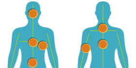
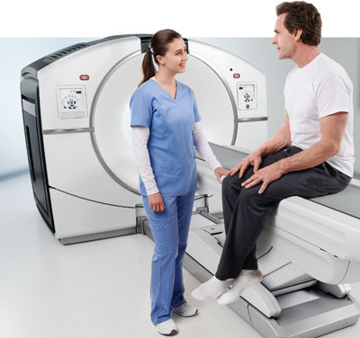 A PET (Positron Emission Tomography) exam is a cutting-edge molecular imaging test that assesses the functionality of organs and tissues through the use of a radioactive tracer. Element utilizes different types of radioactive tracers tailored to target the cells associated with the specific areas of concern for your physician. This specialized exam is combined with a CT (Computed Tomography) scan for comprehensive disease detection. Unlike conventional imaging tests that focus solely on anatomy, PET technology looks at the chemical and metabolic activity of your body’s cells. The integration of PET/CT technology makes possible the early detection of various conditions, including numerous cancers and brain disorders.
Exam Preparation Instructions - Radiotracer Specific
F-18 FDG for Routine Oncology/Metabolic Evaluation
- Nothing to eat or drink for 4 hours prior to your appointment time
- Patient may drink only a small amount of water to take any necessary medication
- Eat a low carbohydrate/no sugar diet beginning 24 hours prior to exam
- No exercise or strenuous physical activity 24 hours prior to exam
- No alcohol or smoking 12 hours prior to exam
- Allow 2 hours at our facility for your PET/CT scan
F-18 PSMA positive lesions in prostate cancer patients with suspected metastasis and candidates for initial definite therapy and/or suspected recurrence based on elevated PSA levels
- No prep required for this exam
- Stay well hydrated
F-18 Fluciclovine (Axumin) Radiotracer for Prostate Cancer Recurrence
- No strenuous activity 24 hours prior to exam
- Nothing to eat or drink 4 hours prior to exam
Cu64 Dotatate (Detectnet) Radiotracer for Neuroendocrine Tumor
- Drink plenty of water the day before and the day of the exam
- Fasting is not required
- Patients on LONG-ACTING SOMATOSTATIN treatment must schedule exam prior to next dose
- Patients on SHORT-ACTING SOMATOSTATIN treatment should discontinue 48 hours prior to exam
F-18 Fluoroestradiol (Cerianna) Radiotracer for Evaluation of ER+ Lesions in Recurrent or Metastatic Breast Cancer
- No prep required for this exam
F-18 Florbetaben Neuraceq Radiotracer for Evaluation of Beta Amyloid Plaque/Alzheimer’s Dementia
- No prep required for this exam
Hysterosonogram
Exam Explanation
Hysterosonography also called Sonohysterography or Saline Infusion Sonography, is a minimally invasive ultrasound exam that provides images of the inside of the uterus to help diagnose the cause of abnormal vaginal bleeding.
Exam Preparation
You should inform your doctor or technologist of any medications being taken and if you have any allergies. You must inform the doctor if you are or might be pregnant. The exam is best performed between days seven and ten of your cycle (if you are pre-menopausal) to minimize the chance that you have ovulated or are already pregnant. This procedure should not be performed if you are pregnant and/or if you have an active inflammatory condition. Unlike hysterosalpingogram (or HSG) this test will not determine if the fallopian tubes are open or blocked.
During the Exam
The procedure is like a gynecological exam. You will be asked to remove your clothes below the waist and change into a gown. You may wish to wear a two-piece outfit that day. You will be asked to empty your bladder. You will lie on your back on the exam table. A transducer is inserted into your vagina to view your uterus and ovaries. A catheter is then inserted into the cervix, and a sterile saline solution is injected into the catheter. Ultrasound images are taken during the injection, allowing the evaluation of the uterine lining. This is approximately a 30 minute exam.
Fast Breast MRI
What is a Fast Breast MRI?
A Fast Breast MRI is an abbreviated version of a traditional Breast MRI, which means fewer images are captured resulting in a shorter time frame for the exam.
This is a screening exam that is available to all patients, and it is a valuable supplement to mammography for patients who are above-average risk. Your Element Medical Imaging radiologist may recommend a Fast Breast MRI after interpreting the findings of your screening mammogram.
This self-pay exam requires a physician referral, and the cost is only $450.00.
What to expect the day of the exam
The entire appointment will take about 30 minutes. This exam should be scheduled 7-10 days after the start of your menstrual cycle. Correct timing is important to minimize false positive findings that can occur due to hormonal influence on the breast tissue. A Fast Breast MRI does not require your breasts to be compressed, so you should not experience discomfort.
When you arrive, you will be asked to complete paperwork regarding your history and symptoms. We will escort you into a private dressing room where you can change into a gown and remove all jewelry, since these items contain metal, which disturbs MRI signals. A female technologist will position you for the scan. During the exam, you will lie on your stomach with your arms up over your head, and you will enter the machine headfirst. Avoid eating a large meal prior to the exam. Most patients receive an injection of contrast material called gadolinium during the exam through an intravenous injection.
You will be asked to lie very still, relax and breathe normally. There are typically no side effects during or after MRI, so you can resume normal activities as soon as your exam is over. It is very important that any prior breast films (mammograms, ultrasound or MRI) be made available to the radiologist for comparison during the interpretation of your MRI scan. If you have had these at a facility other than Element Medical Imaging please bring them with you on the day of your appointment. If you have any of the items listed below, please call 913.469.8998 so we can make arrangements for you before your appointment. Many of these items are contraindications to having an MRI as they are not compatible with the magnetic field present around all MRI machines.
- Cardiac pacemaker
- Artificial heart valve prostheses
- Aneurysm clips
- Eye implants or metal ear implants or any metal implants activated electronically, magnetically or mechanically.
- Copper 7 IUD
- Shrapnel or non-removed bullet
- Pregnancy
- Weight over 350 lbs.
- Claustrophobia
- Any metal puncture(s) or fragment(s) in eye
DEXA (Bone Density)
DEXA (Dual Energy X-ray Absorptiometry) measures bone density. A DEXA scan is safe and painless. It typically takes 10-20 minutes, and minimal radiation is used to determine the bone density of the spine, hip or wrist.
Its results are reproducible, so measurements can be taken over time to show the progression of disease or improvement in bone density with treatment.